- 800.525.2690
- [email protected]
- Mon - Fri: 8:00 - 4:30
New Examination & Earwax Removal Technology for Industrial Safety
Smart technology and micro-cameras make video otoscopes more portable and affordable.

By: By: Robert M. Traynor and Garry G. Gordon
Article first published in Workplace Material Handling and Safety magazine.
Today’s hearing conservation programs require a visual inspection of the ear canal and eardrum prior to hearing testing for fitting with hearing protection devices. This is especially important in programs that use in-the-ear or custom hearing protection devices.
As the workers introduce and remove these insert earplugs from their ears, they typically move the earwax further into the ear canal, and, thus, it is easy to create significant wax buildups or impactions. Therefore, earwax becomes an important obstacle that must be ruled out in the beginning of the hearing examination or an earplug fitting.
What Is Earwax?
Earwax, termed cerumen by professionals, is a naturally occurring substance that cleans, protects, and lubricates the ear canal. While beneficial and usually harmless, ear canal blockages can lead to several frustrating conditions such as hearing loss, tinnitus, fullness, itching, odor, cough, and even significant ear pain.
In severe cases, an earwax impaction can create a totally curable 40 dB or more hearing loss creating difficulty hearing in the affected ear. The presence of impacted earwax can also have significant implications for psychological and emotional health, as well as communication and social functioning.1
Earwax is generated by the combined secretions of the sebaceous glands producing a substance called sebum and apocrine, or sweat glands, within the outer two-thirds of the ear canal. When mixed with the ear canal’s dead skin it becomes what we know as earwax. Its composition can be soft or hard, but there is a self-cleaning mechanism that takes care of most of the earwax that is generated.
This self-cleaning system is part of the ear canal’s generation of new skin that lines the canal. As the skin naturally moves toward the opening of the ear canal, the earwax typically falls out or washes away.
How Is Earwax Removed?
According to UCLA Health2, approximately 12 million Americans spend $51 million each year seeking treatment from health care professionals for earwax issues. In the U.S., professional earwax removal has been traditionally part of family medicine, and more recently, audiologists, unless it is impacted and completely blocking the ear canal. These cases are usually referred to otolaryngologists (physicians specializing in ear, nose, throat) where a sophisticated irrigation procedure is completed without damage to the auditory system.
Health care professionals may provide instructions to safely remove earwax at home, but the following home remedies are not recommended:
- – Slimy ear drops that, after letting them work for a time, are irrigated with a bulb syringe, and often drizzle down the neck.
- – The use of the squirt gun that irrigates the ear. It sometimes removes the earwax and sometimes just makes a mess.
- – Q-tips are a popular method of earwax removal. Unfortunately, using a Q-tip pushes the wax further down into the ear canal creating an impaction.
- – There are also those that put candles into the ear canal and light them thinking that this will remove the earwax, however, it does not. This candling process has been called “the triumph of ignorance over science.”
When there is some disruption of the normal self-cleaning mechanism, such as those presented above, the earwax may need to be removed by professionals, such as physicians or audiologists.
How Are Ears Examined?
For decades, the standard device for ear examination has been the otoscope. This device directs a beam of light through a speculum into the ear allowing observation of the ear canal and eardrum. Although the positioning of the scope is cumbersome, the light is often lacking and the visual field is limited, it has been considered the most affordable best practice for ear examination.
With the introduction of micro cameras in recent years, professionals conducting ear examinations have now integrated a new best practice standard for ear examination or earwax removal which is the use of the video otoscope.
Video Otoscopes: Video otoscopy provides a direct and clear view of the ear canal and eardrum. For hearing conservation managers, however, the costs of $8,000-10,000 or more have been prohibitive. Portability has also been a factor, as these instruments are rather large and cumbersome for many itinerant hearing conservation providers.
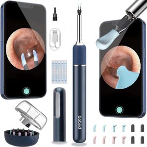
Digital Otoscopes: Using a software application, the video camera in the tip of the digital otoscope is paired with a smart device, such as a smartphone or tablet, enabling the device to capture images and videos of the external ear canal and ear drum. These scopes can often be connected via Bluetooth to a larger monitor to view photos and/or videos during examinations, otoblock insertions or earwax removal procedures.
Due to this technology, the costs have now been drastically reduced to about $125 or so for a high-quality video otoscope. Portability is now facilitated by the use of either an Android or an iPhone (iOS) smartphone or iPad, for easy internet connection and high-quality screens for ear canal and eardrum visibility almost anywhere.

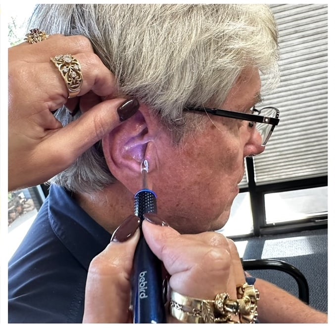
Figure 1: The EAR/Bebird Note 9 is a rechargeable digital otoscope, with a 10 megapixel video camera and software application that is compatible with either Android or iPhone/iPad (iOS) devices. The system includes tips and instruments for ear canal examinations and earwax removal.
Hearing Conservation Applications
Digital otoscopes can be a more affordable option for use in industrial hearing health care to check that the ear canal is clear for earplug use, placement of otoblocks for custom earplugs, or to clear the patient for a hearing test.
For example, one manufacturer, EAR/Bebird offers affordable, rechargeable video otoscopes for use in industrial hearing health care, and personal use. The instruments use an internet connection, a software application and a smartphone or tablet. The system allows for easy ear canal observations for both the examiner and the patient to see the results.
For the examiner, the camera offers a high-resolution check that ensures the ear canal is clear for earplug use, placement of otoblocks for custom earplugs, or to clear a patient for a hearing test. These instruments are compatible with either Android or iPhone/iPad (IOS) applications and present the correct orientation for either right or left ears (Figure 1).
Digital otoscopes can also be used by hearing conservation program examiners, and instruments are available for personal use to assist with ear canal and eardrum visualization and wax removal.
These instruments offer a unique capability to the hearing conservation manager in that they may be used for examination, but also for education and training in ear hygiene. During the education process, employees who wear hearing protection should be informed about their options when excess earwax or impaction is observed.
